Electrical changes during muscular contraction
When the muscle is stimulated, electrical changes occur before onset of mechanical changes. Usually the electrical events in a muscle (or any living tissue) are measured by using a Cathode Ray Oscilloscope. Nowadays, sophisti cated electronic equipments like computerized polygraph are available to record and analyze the electrical activities of any tissue.
RESTING MEMBRANE POTENTIAL
The potential difference between inside and outside of the cell under resting condition is known as resting membrane potential.
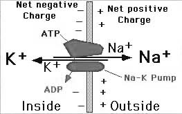
Fig.2. Resting membrane potential.
When two electrodes are connected to a cathode ray oscilloscope through a suitable amplifier and placed over the surface of the muscle fiber, there is no potential difference. There is zero potential difference. But, if one of the electrodes is inserted into the interior of the muscle fiber, potential difference is observed across the sarcolemma (cell membrane). There is negativity inside the muscle fiber in relation to the outside. This potential difference is constant and is called resting membrane potential. The condition of the muscle during resting membrane potential is called polarized state. In human skeletal muscle, the resting membrane potential is -90 mV.
ACTION POTENTIAL
When the muscle is stimulated, a series of changes occur in the membrane potential, which is called action potential. The action potential occurs in two phases.
1.Depolarization and
2.Repolarization.
Depolarization
When the impulse reaches the muscle, the polarized condition (-90 mV) is altered, i.e. the resting membrane potential is abolished. The interior of the muscle becomes positive and outside becomes negative. This condition is called depolarization. With other words, depolarisation is membrane potentials difference decreasing.
Repolarization
Within a short time, the muscle obtains the resting membrane potential once again. Interior of the muscle becomes negative and outside becomes positive. So, the polarized state of the muscle is re-established. This process is called repolarization. So, it is potentials difference restoration.
ACTION POTENTIAL CURVE
Resting Membrane Potential
The resting membrane potential is recorded as a straight baseline at -90 mV .
Stimulus Artifact(local potential)
When a stimulus is applied, there is a slight irregular deflection of baseline for a very short period. This is called stimulus artifact.
|
|
|
Latent Period
The stimulus artifact is followed by a short period without any change. This period is called latent period, which is about 0.5 to 1 millisecond.
Firing Level or Critical Depolarization Level
Depolarization starts after the latent period. Initially, it is very slow. After the initial slow depolarization up to -15 mV, the rate of depolarization increases suddenly. The point at which, the rate of depolarization increases is called firing level.
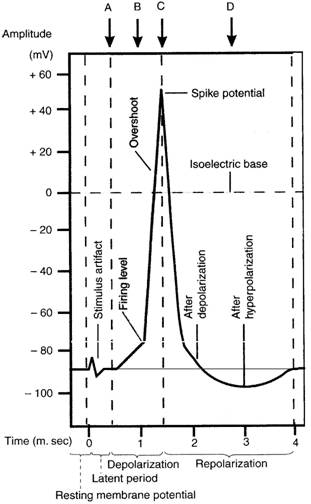
Fig. 3. Action potential in a skeletal muscle.
Overshoot
From firing level, the curve reaches the isoelectric potential (zero potential) rapidly and then overshoots the zero line up to +55 mV.
After Depolarization or Negative after Potential
The rapid fall in spike potential is followed by a slow repolarization process. This is called after-depolarization, trace depolarisation or negative after potential. The duration of this is 2 to 4 milliseconds.
Дата добавления: 2018-02-15; просмотров: 1489; Мы поможем в написании вашей работы! |

Мы поможем в написании ваших работ!